


MIYOLI P21
5D ultrsound that you can afford
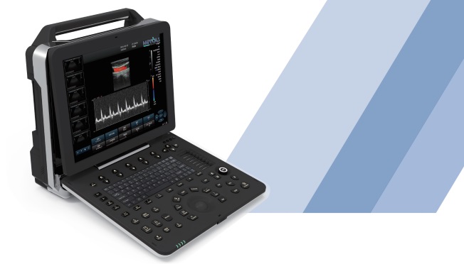
MIYOLI P21 Elite equipped with a high-resolution medical display, adopts multi-beam parallel technology and sub-array element transducers, and its superior image clarity perfectly meets the needs of women‘s health care. At the same time, it relies on the realistic RealSkin 5D ultrasound technology and abundant measurement packages to better product women’s health.
What is HD Live (5d) Technology?
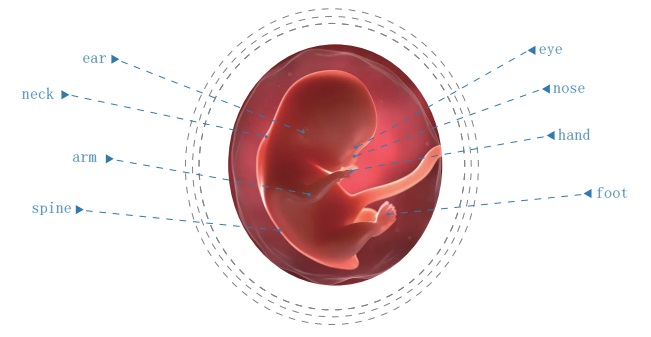
ultrasounds with 3D/4D imaging have be around for years, but the HD is (5D) ultrasound technology uses a unique and moveable light source inside the probe that not only provides both 3D and 4D imaging, but “lights up” the bay, making its possible for-soon-to-be-parents to see to the facial expression of their child, and even watch their bay yawn, wink and smile.
An HD live (5D) ultrasounds provides impeccable views of your providing better depth perception. We can position of the light and more clearly see the baby’s lips, nose and eye lids.
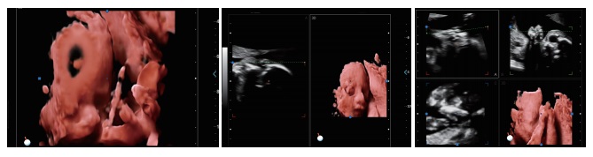
Real skin Rendering
Real skin rendering adopts 4D ultrasonic images plus spatial dimension parameters to obtain more three-dimen- sional and realistic 4D images, which surpasses most of the limitations of traditional gray-scale ultrasound.
Smooth Work flow
Easy operation process, quick response to diagnostic needs, can easily deal with complex situations in mobile diagnosis
- High resolution medical 15. 6 inch display
- The display is adjustable 0 to 30
- Hard disk dynamic and static image storage, real-time sharing
- Spectrum envelope function
- Built-in large capacity lithium battery (detachable)
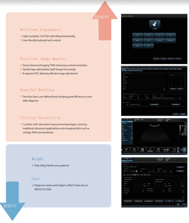
The multi-purpose user presents, comprehensive measurement and report system,built in easy view image archive system, quick image storage/retrieve/transfer, one button direct print, makes the complete workflow better then what you can use of.
Micron Imaging Technology
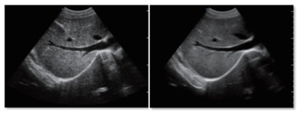
Micron imaging technology, real-time tracking of specic signals at the edges of dierent tissues, to achieve edge enhancement, and monitor each pixel at the same time; optimize the internal signal of the organization and perfectly integrate the edge information and the internal pixel information of the organization to restore the real and delicate, excellent level contrast Two-dimensional image.
Harmonic Imaging Technology (THI)
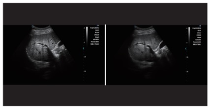
It improves image clarity by improving tissue contrast resolution, spatial resolution, and eliminating near-eld artifacts. It is mainly used for the diagnosis of cardiovascular and abdominal diseases. It plays an important role in evaluating the lesion area and boundary division of patients with imaging diculties. The technology has been fully approved by clinicians. Harmonic technology retains the second harmonic signal to the greatest extent on the basis of removing the fundamental signal, which increases the signal strength by more than 30% compared with the traditional signal processing, reduces noise and artifacts, and improves the contrast resolution of tissue images.
Trapezoid Imaging
Trapezoid imaging is a kind of expanded imaging, which is transformed into a trapezoid on the basis of the original rectangle, and the left and right sides are expanded to a certain extent, achieving a wider eld of view. The principle of ultrasound imaging is to scan the human body with ultrasonic sound beams, and obtain images of internal organs by receiving and processing the reected signals.
Automatic spectrum tracking measurement technolo
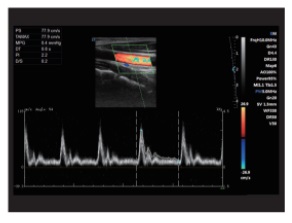
Ultrasound Doppler technology is used in the ultrasound system for examining the heart and arteries and veins. It is necessary to extract relevant parameters from the Doppler spectrogram to evaluate the hemodynamic status of the heart and blood vessels. The disadvantage of manual detection is that the operator's marking of the peak velocity is relatively monotonous and time-consuming, with poor repeatability and low estimation accuracy; and during the detection, in order to mark the peak velocity, the operator needs to interrupt the acquisition of Doppler signals, which makes it impossible to estimate in real time.
This host contains an automatic envelope detection module, which can automatically track the time-related changes of the peak blood ow velocity and average velocity, and display them in real time on the Doppler spectrogram.


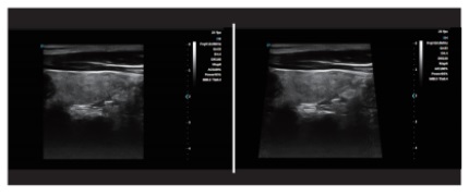
Sub-Array Technology
The dedicated high-density probe adopts a brand-new array design technology and a unique sub-array element technology. The second cutting of independent wafers can completely control the entire process of wafer vibration, thereby reducing side lobe artifacts and enhancing the ne resolution of tissues. The boundary between adjacent strong echo reectors is sharper. It fully demonstrates the high-resolution images brought by the high-density probe, perfectly presents the image details, and increases the accuracy of clinical diagnosis.